
Molecularly Engineered Scaffolds for Accelerated Wound Healing and Scar Reduction
- Dr.N. Thirumalaivasan
- Dec 24, 2025
- 3 min read

Updated: Jan 29
Wound Healing Scaffold Technologies
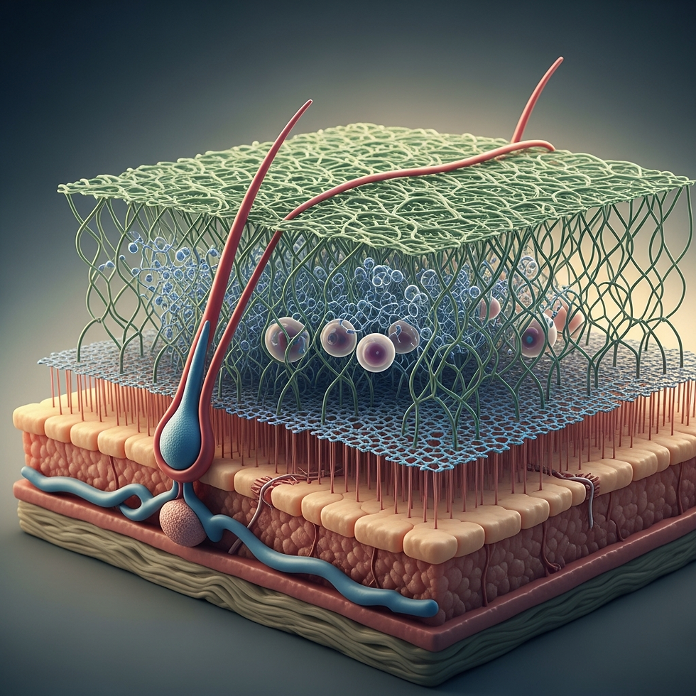
Wound healing scaffolds represent a core innovation in regenerative medicine, functioning as three-dimensional biomaterial systems engineered to recapitulate the physicochemical and biological functions of the native extracellular matrix (ECM). These platforms provide mechanical stabilization while actively orchestrating cellular recruitment, angiogenesis, matrix remodeling, and controlled therapeutic delivery, thereby accelerating tissue regeneration and minimizing fibrotic scarring.
Mechanistic Role in Tissue Repair
Scaffolds promote wound repair by establishing a permissive microenvironment that regulates cell adhesion, proliferation, migration, and lineage commitment. Their hierarchical porous networks enable efficient mass transport of oxygen, nutrients, and metabolites, sustaining fibroblast viability and endothelial cell infiltration. Scaffold architecture further supports granulation tissue formation and spatiotemporal coordination of collagen deposition, facilitating organized tissue remodeling and restoration of skin integrity.
Functional Performance and Design Criteria
An ideal scaffold exhibits high biocompatibility, immunological tolerance, and predictable biodegradation synchronized with tissue regeneration rates. Tunable mechanical properties ensure compatibility with skin elasticity and wound biomechanics, while interconnected pore systems enhance vascularization and tissue ingrowth. Functionalization with growth factors, antimicrobial agents, or gene vectors enables targeted molecular modulation of inflammation, angiogenesis, and ECM synthesis.
Molecular-Level Signaling and Regenerative Mechanisms
At the molecular scale, wound healing scaffolds actively regulate intracellular signaling pathways that govern inflammation resolution, cell migration, and matrix regeneration. ECM-mimetic biochemical cues interact with cell-surface integrins to activate FAK (Focal Adhesion Kinase) and PI3K/Akt signaling, promoting cytoskeletal organization, survival, and proliferative responses.
Scaffold-mediated release of growth factors such as VEGF, PDGF, and TGF-β stimulates angiogenesis, fibroblast activation, and collagen biosynthesis through MAPK/ERK and Smad-dependent pathways. Additionally, modulation of matrix metalloproteinases (e.g., MMP-9) and their inhibitors helps rebalance proteolytic activity, preventing chronic inflammation and ECM degradation in non-healing wounds.
Advanced bioactive scaffolds can influence immune cell polarization by promoting a shift from pro-inflammatory M1 macrophages to regenerative M2 phenotypes, thereby enhancing cytokine profiles favorable to tissue repair (e.g., IL-10, Arg-1). Emerging nano-engineered scaffolds further enable controlled microRNA and gene delivery, allowing fine-tuned regulation of cell differentiation, angiogenic signaling, and fibrosis suppression at the transcriptional level.
Stage-Specific Modulation of Wound Healing
Inflammatory Phase: Controlled release of anti-inflammatory agents and protease inhibitors reduces excessive cytokine signaling and oxidative stress.
Proliferative Phase: Scaffolds enhance fibroblast expansion, endothelial sprouting, and granulation tissue formation through angiogenic and mitogenic signaling.
Remodeling Phase: Gradual scaffold degradation supports collagen realignment and ECM maturation, favoring functional tissue regeneration over scar formation.
Emerging Research Directions
Current research emphasizes smart, stimuli-responsive scaffolds capable of responding to pH, temperature, or enzymatic activity to regulate drug release and immune responses. Integration of nanomaterials, metal-based antimicrobial agents, stem-cell supportive matrices, and bioelectronic sensing platforms is driving the transition from passive wound dressings to active, adaptive regenerative systems. Ongoing translational efforts aim to achieve scalable manufacturing, regulatory compliance, and personalized wound care solutions.
Table: Scaffold Materials, Functional Roles, and Clinical Relevance
Scaffold Material / Platform | Key Biological Functions | Advantages | Clinical / Translational Relevance | |
Collagen | ECM mimicry, fibroblast adhesion, angiogenesis | High biocompatibility, natural bioactivity | Chronic wound dressings, skin graft substitutes | |
Chitosan | Antimicrobial activity, hemostasis, cell adhesion | Biodegradable, infection-resistant | Burn wounds, diabetic ulcer treatment | |
Silk Fibroin | Mechanical strength, controlled degradation | High tensile stability, low immunogenicity | Advanced wound dressings, tissue-engineered skin | |
Polycaprolactone (PCL) | Structural support, slow biodegradation | High durability, tunable mechanics | Long-term scaffolds for deep wounds | |
Polyurethane | Elasticity, moisture retention | Flexible, mechanically resilient | Elastic wound coverings, chronic wound care | |
Decellularized ECM | Native biochemical signaling, integrin activation | High biomimicry, enhanced tissue integration | Skin regeneration, reconstructive surgery | |
Hydrogels | Moisture retention, drug delivery | Injectable, stimuli-responsive | Smart wound dressings, controlled drug release | |
Electrospun Nanofibers | Cell guidance, high surface area | ECM-like fiber architecture | Regenerative wound patches, angiogenic scaffolds | |
Nanocomposite Scaffolds | Antimicrobial, immunomodulatory, gene delivery | Multifunctional, smart response | Next-generation personalized wound therapies |


Comments