
Rational Design of Subcellular Fluorescent Biomarker
- Dr.N. Thirumalaivasan
- Dec 24, 2025
- 2 min read

Updated: Dec 26, 2025
Our laboratory specializes in the rational design of subcellular fluorescent biomarkers for high-resolution live-cell imaging. By integrating molecular engineering and nanomaterial design, we develop organelle-targeted, stimuli-responsive probes to visualize intracellular processes for applications in cell biology, cancer diagnostics, and precision nanomedicine.
Research Overview
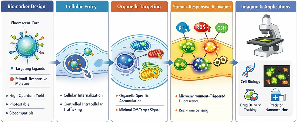
Our laboratory is dedicated to the rational design and preparation of advanced subcellular fluorescent biomarkers for precise visualization of intracellular processes with high spatial and temporal resolution. The central objective of this research is to create organelle-specific fluorescent probes that selectively localize within key subcellular compartments—nucleus, mitochondria, lysosomes, endoplasmic reticulum, and Golgi apparatus—thereby enabling accurate mapping of cellular structure and function.
Molecular and Nanomaterial Design Strategy
Our approach integrates rational molecular engineering with nanomaterial-based design principles. By systematically tuning surface chemistry, molecular charge distribution, hydrophilicity, and organelle-targeting ligands, we achieve controlled cellular internalization, directed intracellular trafficking, and selective accumulation within desired subcellular regions. This design strategy ensures predictable localization behavior and minimizes off-target background signals.
Optimized Optical and Biological Performance
A major focus is placed on engineering fluorescent biomarkers with:
High fluorescence quantum yield
Superior photostability
Low background interference
Minimal cytotoxicity
Excellent biocompatibility
These features enable robust live-cell imaging and long-term intracellular tracking, ensuring reliable performance in complex biological environments.
Stimuli-Responsive and Functional Imaging
A distinctive strength of our research lies in the development of stimuli-responsive subcellular fluorescent biomarkers. These smart probes dynamically respond to local microenvironmental cues, including:
pH fluctuations
Redox imbalance
Reactive oxygen species (ROS)
Glutathione (GSH) levels
Enzymatic activity
Such responsiveness allows real-time monitoring of organelle health, oxidative stress, metabolic dynamics, and disease-associated cellular transformations, providing functional insights that extend beyond static imaging.
Advanced Imaging Validation
Comprehensive validation is performed using state-of-the-art fluorescence imaging platforms, including confocal and super-resolution microscopy. These techniques are employed to rigorously assess targeting precision, intracellular stability, signal fidelity, and temporal responsiveness, ensuring translational relevance and reproducibility.
Translational Impact and Applications
The outcomes of this research contribute enabling technologies across multiple domains, including:
Fundamental cell biology
Cancer diagnostics
Intracellular drug delivery tracking
Precision nanomedicine
By offering powerful tools to visualize and interrogate complex subcellular events, this work supports next-generation strategies for diagnosis, therapeutic monitoring, and mechanistic understanding of disease at the cellular level.

Comments